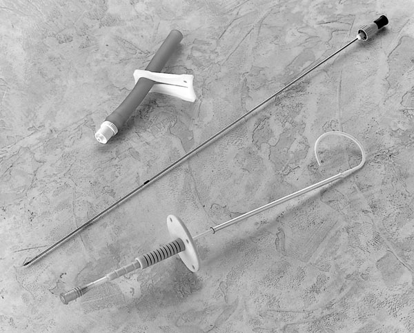
Ideally ascitic procedures should be ultrasound guided
Indications for ascitic drain insertion (therapeutic paracentesis)
- Refractory ascites secondary to portal hypertension (usually in liver cirrhosis)
- Palliation in malignant ascites
- Respiratory embarrassment (secondary to diaphragmatic ‘splinting’)
|
Equipment required for ascitic drain insertion (therapeutic paracentesis)
- Ultrasound and ultrasound operator
- Dressing trolley & sharps bin
- Sterile field
- Sterile dressing pack
- Sterile gloves
- 2% Chlorhexadine swabs
- Analgesia
- 10mls of 1% or 2% Lidocaine
- Orange (25G) needle (x1)
- Green (19G) needle (x1)
- 10ml Syringe (x1)
- 20ml Syringe (x1)
- Scalpel
- Cannula dressing (x2)
- Paracentesis catheter (Safe-T-centesis®, Bonnano or similar 18G drain)
- Urinary catheter bag (or similar)
- Blood culture bottles
- 20% Human Albumin Solution (HAS)
|
Contraindications to ascitic drain insertion (therapeutic paracentesis)
- Local infection
- Cautions – but not contraindications
- Coagulopathy (INR>2.0)
- Attempt to correct INR to <1.5 if possible.
- Platelets<50
- Thrombocytopenia and coagulopathy is often present in liver disease and though it is a caution, it not a contraindication to paracentesis or drainage
- The incidence of clinically significant bleeding is low; routine FFP or platelets is not indicated
- Pregnancy
- Organomegaly
- Obstruction/ileus
- Distended bladder
- Abdominal adhesions
|
Potential complications of ascitic drain insertion (therapeutic paracentesis)
- Perforation of viscus or vessels causing haemorrhage (abdominal wall haematoma has been reported in up to 2% in case series)
- Intra-vascular volume depletion (hypotension) & renal impairment
- Exacerbation of hepatic encephalopathy
|
Pre-procedure
- Consent patient
- Infection, bleeding, pain, failure, damage to surrounding structures (especially bowel perforation), leakage
- Ultrasound to confirm fluid and insertion sight (see ascitic tap pages)
- Set up sterile trolley
|
Procedure for ascitic drain insertion (therapeutic paracentesis)
- Position the patient supine in the bed with their head resting on a pillow.
- Select an appropriate point on the abdominal wall in the right or left lower quadrant, lateral to the rectus sheath. If a suitable site cannot be found with palpation and percussion consider using ultrasound to mark a spot.
- Clean the site and surrounding area with 2% Chlorhexadine and apply a sterile drape.
- Anaesthetise the skin with Lidocaine using the orange needle. Ensure you raise a large bleb as the drain perforating the skin will be the most painful part of the procedure.
- Anaesthetise deeper tissues using the green needle, aspirating as you insert the needle to ensure you are not in a vessel before infiltrating with lidocaine. Use a maximum of 10mls of Lidocaine.
- Take the Bonanno catheter and advance needle to tip of catheter, thus straightening it out
- Insert the paracentesis catheter using a ‘Z’ track
- Perforate the skin perpendicularly, and then advance obliquely in the sub-cutaneous tissue for 1-2cm before returning to a perpendicular position to puncture the peritoneal cavity.
- Gradually advance the catheter into the peritoneal space.
- Once you have inserted the catheter to the equivalent length of the green needle where fluid was first aspirated, start to pull the needle back slowly whilst advancing the catheter.
- Do not pull the needle back too far as it is needed for stability, but equally do not push the needle too far into the peritoneal cavity.
- Advance catheter to the hilt and completely remove needle.
- Fix with two sterile cannula dressings.
- Affix the drainage bag and leave on free drainage after obtaining the required samples.
- Microbiology
- Microscopy, culture & sensitivities (be explicit if yeast or mycobacterium suspected)
- Culture in blood culture bottles inoculated at the bedside
- Haematology
- Automated WCC count (send EDTA sample)
- Biochemistry
- Albumin, Protein, LDH, Glucose
- Remember to send a serum albumin, LDH and glucose at the same time (or at least from the same day).
- Special tests: Fluid amylase, Triglycerides, Bilirubin
- Cytology
- Remove drain after 6 hours if cirrhosis
- This is due to the high risk of peritonitis
- Drains for malignant effusions can be left in for longer but the risk of peritonitis still exists
|
Post-procedure
- Monitor Pulse, BP and Respirations
- 15 minutes for 1 hour; 30 minutes for 1 hour; And hourly for 4 hours
- Measure and record drain and urine output
- Observe for signs of shock or acute haemorrhage
- In patients with liver cirrhosis do not leave drain in for more than 6 hours
- In patients with cirrhosis consider infusing human albumin solution for every litre drained – liaise with gastroenterology for advice if needed
- In portal hypertensive ascites order 20% Human Albumin Solution from the blood bank. Generally 100mls should be infused for each 2000mls of ascites drained.
- Volume replacement is not routinely required for malignant ascites unless the patient becomes hypotensive during drainage (but suggest 250ml colloid fluid challenge if required).Send fluid for urgent cell count, MC&S, LDH, protein and cytology
- Send paired LDH and protein serum samples
- Consider antibiotic cover if SBP is suspected
- Refer to your trust policy
- Co-amoxiclav and Tazocin are commonly used
|
In the event of failure
- Stop procedure
- Seek senior help
- Re-review imaging and patient with a senior colleague to ensure presence of fluid
- Consider further imaging or ascitic drain insertion in radiology
|
Top Tips for ascitic drain insertion (therapeutic paracentesis)
- Inserting a Bonanno catheter requires a similar motion to cannula insertion, it is important not to advance the needle too far but you need to ensure the catheter is passing into the peritoneum without kinking.
- A Bonanno catheter is actually a form of suprapubic catheter. When ascitic drains are inserted in the radiology department, they will use pigtail catheters
- In patients with a thick abdominal wall a spinal needle can be used to infiltrate anaesthetic and check position.
- If you aspirate blood when infiltrating an anaesthetic; stop, withdraw your needle, change position by 1-2cm and try again.
- If your patient becomes more hypotensive whilst being drained then temporarily clamp the drain and infuse colloid fluid iv (e.g 20% Human Albumin Solution or Gelofusin®).
|


