


Nose Examination
How to examine the nose for doctors, medical student finals, OSCEs and MRCP PACES
Introduction
- Wash hands and apply alcohol gel
- Introduce self
- Explain what you are going to do
- Obtain verbal consent
- Ask if any part of the nose is painful to the touch
Equipment Required
- Thudicum’s speculum or flexible nasendoscopy (see below)
- Rhinoscopy
- A torch (ideally a head torch)
- An auroscope, with the largest aural speculum
- Flexible and rigid endoscopy often with appropriate radiological imaging
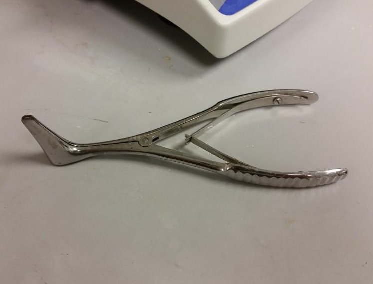
Thudicum’s speculum
General Inspection
- Begin with an overall inspection of the nose
- Evaluate the size and shape
- Lift the lip of the nose with a gloved thumb to obtain a better view and observe the colour and texture of the skin
- Butterfly rash over the nasal bridge
- Malar rash in systemic lupus erythematosus
- Thickening and coarseness seen in hypothyroidism
- A large bulbous nose with a coarse “orange skin” like appearance suggests a rhinophyma

Rhinophyma
- Check for any telangectasia on the face or hands
- Inspect from the side for any deformity of the nasal bridge
- Look underneath the nose to check the nostrils for symmetry and narrowing
- Determine if the nasal septum has become dislocated from the collumella partly or even completely blocking the nasal airway

Deviated nasal septum

Septal haematoma
Palpation
- Before palpating the nose, ask the patient to if there is any pain or tenderness
- If trauma has taken place, then gently feel for:
- Mobile nasal bones
- A fracture line
- A step or depression (which can occur if the nasal bone has been dislocated inwards)
- If trauma has led to a septal haematoma (see above) often described as “cherry red” swelling in appearance, then gently pressing the nasal tip is associated with significant discomfort
- It is important to recognise a septal haematoma because if it is not treated with surgical drainage and antibiotics it will often progress to an abscess with loss of the nasal septal cartilage
- This can lead to significant changes to the shape of the nose or rarely cavernous sinus thrombosis
Video on the nasal examination
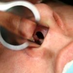
Anterior rhinoscopy
- Start by pushing on the tip of the nose in an upwards direction and look in to the anterior nares
- Deliberately and systematically inspect the roof of the nose and along the floor of the nose.
- Make a note of any particular dryness but remember that as the patient ages, the tendency is for the nose to become gradually dry.
- Mucous is found quite infrequently in the nose and its colour and thickness may help identify its cause.
- Clear and watery mucous: nasal allergy or an upper respiratory tract infection
- Yellow or green mucous: infection (viral or bacterial) or stasis
- Mucopurulent discharge is unilateral in an adult is likely to be a sinus infection but especially in a child consider the possibility of a foreign body.
- Thick white mucus: chronic rhinosinusitis.
- A fractured cartilaginous nasal septum due to trauma and developed a septal abscess, There may be a perforation of the nasal septum (see below).
- Usually these are benign following trauma
- Also Below is a clinical picture obtained with an endoscope showing a nasal polyp emerging from between the middle and inferior turbinate and demonstrating the difference in appearance between the turbinate and a polyp
- Epistaxis is a clinical term used to describe bleeding from the nose and is the commonest nasal emergency
- Most epistaxis, arises from Little’s area at the anterior end of the nasal septum
- This region of the nose has a rich blood supply from the anterior ethmoidal artery, the sphenopalatine artery and the septal branch of the superior labial artery
- Video below shows silver nitrate (75%) being applied to a vessel in Little’s area to control an epistaxis
 A Perforation of the nasal septum
 Polyp emerging from the turbinate
Testing of the Olfactory nerve
- Loss of the sense of smell is termed anosmia
- True anosmia can be identified using the ‘Smell Identification Test’
- This is a self administered scratch and sniff test consisting of four booklets, each booklet containing ten stimuli for smell
Testing nasal cilial function
- Normal cilia move mucus from the front to the back of the nose. The common cold can affect cilia beat frequency and the usual coordinated action of the cilia leading to stasis of mucus
- Primary ciliary dyskinesias are a group of conditions where the cilia fail to move in a coordinated fashion leading to mucus stasis
- If a primary cilial dyskinesis is suspected then the saccharine transit time can be measured
- This can be done by placing a small fragment of saccharine on the anterior end of the inferior turbinate and timing how long it takes the patient to taste it. If the patient tastes the sweetness this is useful to rule out ciliary dyskinesia, though a negative result is of little value
- Other tests include nasal nitric oxide, high speed videomicroscopy (HSVM) and transmission electron microscopy (TEM)
- Often a combination of these tests are used
silver nitrate (75%) being applied to a vessel in Little’s area to control an epistaxis
Conclude the Examination
- Ensure the patient is comfortable
- If your examination suggests a diagnosis incorporating another system (such as the ear or neck) then this should also be examined
- Don’t forget to complete the same hand-washing routine at the end of the clinical examination
Click here to learn about the ear examination and other ENT problems
Perfect revision for medical student finals, OSCES and PACES